Photo by
Yoav Dudkevitch/TPS-IL on 8 March, 2023
New Israeli Research Model Could Shed Light on Intestinal Regeneration
Jerusalem, 18 November, 2026 (TPS-IL) -- Israeli researchers have developed a method for keeping miniature, laboratory-grown human intestinal tissues alive without the growth-factor supplements normally required to sustain them, offering a new tool for studying how the intestine repairs and renews itself.
The breakthrough centers on telocytes, specialized support cells that help maintain intestinal stem cells, which continually replenish the gut’s inner lining. By developing a version of these normally scarce cells that can multiply indefinitely under laboratory conditions, the researchers have created a new way to investigate the intestine’s natural support system.

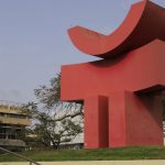
The Hebrew University of Jerusalem campus on Aug. 3, 2023. Photo by Yoav Dudkevitch/TPS-IL
The study, published in the peer-reviewed journal Cellular and Molecular Gastroenterology and Hepatology, was led by Dr. Michal Shoshkes-Carmel and doctoral students Marco Canella and Amal Gharbi at the Institute for Medical Research Israel-Canada at the Hebrew University of Jerusalem’s Medical School. The research was conducted in collaboration with the Organoid Center of Hadassah-Hebrew University Medical Organization.
The intestine constantly renews its inner lining, replacing cells as they wear out. This process depends on intestinal stem cells, which receive essential signals from neighboring cells in a specialized environment known as the stem-cell niche.
Telocytes are an important component of this support system. They form networks beneath the intestinal lining, using long, branching extensions and chemical signals to help maintain stem-cell activity.
Scientists have struggled to study telocytes because they are found in relatively small numbers, are difficult to extract from tissue and multiply poorly under standard laboratory conditions.
A New Research Tool
The Israeli team addressed this challenge by developing an “immortalized telocyte line” — a population of cells modified to multiply continuously under laboratory conditions. The cells retained important features of normal telocytes, including their distinctive extensions, interconnected networks and genes associated with supporting intestinal stem cells.
The researchers then tested whether these cells could sustain intestinal organoids, three-dimensional tissue models often called “mini-guts,” without the growth-factor supplements normally required.
Organoids grown in basic laboratory medium without additional support died within 48 hours. By contrast, organoids cultured alongside the immortalized telocytes survived and continued growing without the supplements. At later stages, they reached sizes comparable to those grown in conventional growth-factor-enriched medium.
The researchers also tested the system using human colon organoids, extending the findings to another type of intestinal tissue model. These miniature tissues failed to survive in basic medium alone but persisted and grew when cultured alongside the telocytes during the observation period.
“Intestinal stem cells depend on essential signals from telocytes: eliminating these support cells or blocking their secretion of Wnt signals disrupts stem cell function. Yet telocytes are exceptionally difficult to grow and study in the laboratory,” Shoshkes-Carmel said.
Wnt signals are molecular messages that help regulate stem-cell activity and tissue renewal.
“By creating an expandable telocyte line, we now have a practical tool for investigating how these cells help sustain the intestinal lining and how their signals may contribute to tissue repair,” she added.
Beyond supporting organoid survival, the telocytes also appeared to influence organoid development. Human colon organoids displayed different structural forms, including rounded and budding shapes, alongside differences in a protein associated with stem-cell activity.
The model does not fully replicate the living intestine. Telocytes grown in isolation lost some of the specialized characteristics associated with their locations in the body, suggesting that signals from surrounding intestinal cells may be necessary to maintain their full identity.
The findings provide researchers with a new laboratory model for investigating how intestinal support cells interact with stem cells and contribute to tissue renewal. The cells are available to researchers upon request, subject to a material transfer agreement.